The Underlying Causes of Ocular Hypertension

Ocular hypertension, also known as high intraocular pressure (IOP), occurs when the pressure within the eyes exceeds the normal range. This condition poses a potential risk for the development of glaucoma, an eye ailment that, if untreated, can result in the loss of vision. To effectively address the risks associated with ocular hypertension and successfully manage its symptoms, it is crucial to comprehend its underlying causes.
Understanding Intraocular Pressure
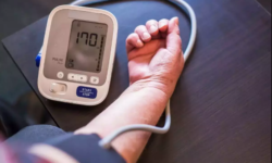
Before we explore the underlying causes of ocular hypertension, it is important to have a clear understanding of intraocular pressure (IOP). Within the eye, there is a fluid called aqueous humor, which serves multiple purposes such as maintaining the eye’s shape, supplying nutrients to the eye tissues, and eliminating waste products. The IOP refers to the pressure exerted by this fluid within the eye.
Key Facts about IOP
| Factor | Description |
|---|---|
| Normal IOP range | 12-22 mmHg |
| Ocular Hypertension | IOP over 22 mmHg |
| Glaucoma | Typically occurs with an IOP over 22 mmHg, though it can also occur within the normal range |
Causes of Ocular Hypertension
Ocular hypertension arises when there is an imbalance in the equilibrium between the creation and drainage of the clear fluid, known as aqueous humor, within the eye. The following are the common factors that contribute to this condition:
Excessive Production of Aqueous Humor
Aqueous humor, a transparent liquid that fills the front chamber of the eye, serves the purpose of nourishment and regulation of intraocular pressure (IOP). However, when the ciliary body, responsible for generating the aqueous humor, starts producing an excessive amount of this fluid, it can cause a rise in IOP. This particular condition, referred to as overproduction of aqueous humor, can stem from various underlying causes and may give rise to severe complications if not promptly addressed.
Causes of Overproduction of Aqueous Humor:
| Causes | Details |
|---|---|
| Idiopathic overproduction | In certain instances, the precise reason for the heightened production of aqueous humor remains elusive. This idiopathic overproduction can manifest without any discernible underlying condition. |
| Inflammatory conditions | Specific inflammatory eye conditions, like uveitis or iritis, can trigger an excessive production of aqueous humor by stimulating the ciliary body. Inflammation within the eye can disrupt the usual regulatory mechanisms, consequently resulting in an overproduction of this fluid. |
| Medications | Certain medications, including corticosteroids, have the potential to stimulate the ciliary body and augment the production of aqueous humor. Prolonged usage of such medications or administration of high doses can be a contributing factor to the overproduction of this fluid. |
| Tumors or neoplasms | In rare instances, tumors or neoplasms located in the eye, particularly in the ciliary body or nearby structures, can disturb the typical regulation of aqueous humor production. This disruption can lead to an abnormal increase in the production of aqueous humor, resulting in overproduction. |
Effects of Overproduction of Aqueous Humor:
The overproduction of aqueous humor can lead to elevated intraocular pressure (IOP), which is a significant risk factor for developing glaucoma. Increased IOP puts pressure on the optic nerve, gradually damaging it and potentially leading to vision loss if left untreated. The effects of overproduction of aqueous humor may include:
- Glaucoma: Chronic elevation of IOP due to overproduction can cause damage to the optic nerve, resulting in glaucoma. Glaucoma is a group of eye conditions characterized by progressive loss of peripheral vision and, if untreated, can lead to blindness;
- Ocular discomfort: The increased fluid can cause discomfort, pain, or a sensation of pressure in the eye;
- Vision changes: Overproduction of aqueous humor can affect the clarity of vision, leading to blurred or distorted vision.
Treatment of Overproduction of Aqueous Humor:
Treating overproduction of aqueous humor typically involves addressing the underlying cause, if known. The primary goal is to regulate the production and outflow of aqueous humor to maintain normal IOP. Treatment options may include:
- Medications: Eye drops or oral medications that reduce aqueous humor production or increase its outflow may be prescribed. These medications may include beta-blockers, prostaglandin analogs, carbonic anhydrase inhibitors, or alpha-agonists;
- Laser therapy: Laser procedures, such as laser trabeculoplasty or laser iridotomy, can help improve the outflow of aqueous humor and reduce IOP;
- Surgical intervention: In some cases, surgical procedures like trabeculectomy or tube shunt implantation may be necessary to manage overproduction of aqueous humor and control IOP.
Regular eye examinations and close monitoring of IOP are essential for early detection and management of overproduction of aqueous humor. Prompt treatment can help prevent vision loss and maintain ocular health. It is crucial to consult an ophthalmologist or eye care specialist for a comprehensive evaluation and personalized treatment plan.
Inadequate Drainage of Aqueous Humor
Aqueous humor is a clear fluid that circulates within the eye, providing nourishment and maintaining intraocular pressure (IOP). However, when the drainage system of the eye, specifically the trabecular meshwork, becomes blocked or inefficient, it can lead to inadequate drainage of aqueous humor. This accumulation of fluid can result in an increased IOP, which can have serious implications for eye health. Understanding the causes and effects of inadequate drainage is crucial for effective management and prevention of complications.
Causes of Inadequate Drainage of Aqueous Humor:
| Causes | Details |
|---|---|
| Primary open-angle glaucoma | The most common cause of inadequate drainage is primary open-angle glaucoma (POAG). In POAG, the trabecular meshwork gradually becomes less effective in draining aqueous humor, leading to increased IOP over time. |
| Secondary glaucoma | Various underlying conditions or factors can contribute to inadequate drainage and secondary glaucoma. These may include eye trauma, certain medications (e.g., corticosteroids), eye inflammation (uveitis), neovascularization (abnormal blood vessel growth), or certain eye anatomical abnormalities. |
| Angle-closure glaucoma | In angle-closure glaucoma, the drainage angle between the iris and the cornea becomes narrow or completely blocked, preventing the normal outflow of aqueous humor. This can cause a sudden increase in IOP and requires immediate medical attention. |
Effects of Inadequate Drainage of Aqueous Humor:
Inadequate drainage of aqueous humor and subsequent elevation of IOP can lead to various eye health complications, including:
- Glaucoma: Chronic elevation of IOP due to inadequate drainage can cause optic nerve damage, resulting in glaucoma. Glaucoma is a progressive eye condition that can lead to irreversible vision loss if left untreated;
- Optic nerve damage: Increased IOP can exert pressure on the optic nerve, causing damage and leading to vision impairment or loss;
- Vision changes: Inadequate drainage can affect the clarity of vision, leading to blurred vision, halos around lights, or decreased visual acuity.
Treatment of Inadequate Drainage of Aqueous Humor:
Managing inadequate drainage of aqueous humor aims to improve the outflow of fluid and regulate IOP. Treatment options may include:
- Medications: Eye drops or oral medications can be prescribed to reduce aqueous humor production or enhance its drainage. These medications may include prostaglandin analogs, beta-blockers, carbonic anhydrase inhibitors, or alpha-agonists;
- Laser procedures: Laser therapy, such as selective laser trabeculoplasty (SLT) or laser peripheral iridotomy (LPI), can help improve drainage by creating openings or enhancing the function of the drainage system;
- Surgical interventions: In cases where medication or laser therapy is insufficient, surgical procedures like trabeculectomy, aqueous shunt implantation, or minimally invasive glaucoma surgery (MIGS) may be considered to create alternative drainage pathways.
Regular monitoring of IOP and routine eye examinations are crucial for early detection and management of inadequate drainage. Timely intervention can help prevent vision loss and preserve ocular health. Consultation with an ophthalmologist or eye care specialist is essential for a comprehensive evaluation and individualized treatment plan.
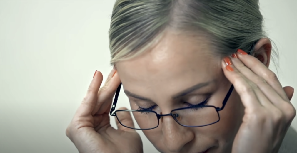
Eye Trauma
Eye trauma refers to injuries sustained by the eye, which can occur due to accidents, sports-related incidents, or other physical trauma. When the eye experiences trauma, it can result in various complications, including an increase in intraocular pressure (IOP). The severity and location of the trauma can determine the extent of IOP elevation and potential damage to the eye’s structures. Understanding the causes, effects, and management of eye trauma-induced increased IOP is crucial for appropriate treatment and prevention of long-term complications.
Causes of Eye Trauma:
| Causes | Details |
|---|---|
| Blunt force trauma | Impact to the eye from a blunt object, such as a punch, ball, or accident-related injury, can cause trauma. The force of impact can lead to inflammation, internal bleeding, or damage to the eye’s structures. |
| Penetrating injuries | Sharp objects, including glass shards, projectiles, or even tools, can penetrate the eye and cause direct damage to the internal structures. Penetrating injuries can result in both immediate and long-term complications. |
| Chemical burns | Exposure to caustic substances, such as acids or alkalis, can cause chemical burns to the eye. These burns can lead to tissue damage, inflammation, and potentially affect the drainage system, leading to increased IOP. |
Effects of Eye Trauma on IOP:
Eye trauma can result in temporary or permanent elevation of IOP due to various mechanisms:
- Inflammation: Trauma to the eye can trigger an inflammatory response, causing swelling and obstruction of the eye’s drainage pathways. This obstruction can impede the normal outflow of aqueous humor, leading to increased IOP;
- Internal bleeding: Trauma-induced bleeding within the eye can increase pressure within the ocular structures, including the anterior chamber. The accumulation of blood can disrupt normal fluid dynamics, affecting the balance of aqueous humor production and drainage;
- Structural damage: Severe trauma can cause damage to the eye’s structures, such as the trabecular meshwork, ciliary body, or drainage angle. Disruption of these essential components can impair the outflow of aqueous humor and contribute to elevated IOP.
Treatment of Eye Trauma and Increased IOP:
The management of eye trauma and increased IOP depends on the extent and severity of the injury. Prompt medical attention is crucial to minimize the risk of complications and preserve vision. Treatment options may include:
- Immediate first aid: Rinse the eye with clean water or saline solution to flush out any foreign particles or chemicals. Avoid rubbing the eye and seek medical help immediately;
- Medications: Eye drops or oral medications may be prescribed to reduce inflammation, control IOP, prevent infection, or manage pain. These medications may include anti-inflammatory drugs, antiglaucoma medications, antibiotics, or analgesics;
- Surgical intervention: In cases of severe trauma or structural damage, surgical procedures may be necessary to repair or reconstruct the eye’s structures. These procedures may include suturing, removal of foreign bodies, vitrectomy (removal of the gel-like substance in the eye), or reconstructive surgery;
- Follow-up care: Regular monitoring of IOP and comprehensive eye examinations are crucial to evaluate the progress of healing and identify any long-term complications. Additional treatments or interventions may be recommended based on the individual’s condition.
It is important to consult an ophthalmologist or seek immediate medical attention in the event of eye trauma. Timely intervention and appropriate management can help minimize the risk of vision loss and maximize the chances of a successful recovery.
Certain Medications
Some medications, particularly steroidal drugs like corticosteroids, can have the side effect of causing an increase in intraocular pressure (IOP). Understanding the potential effects of these medications on IOP is essential for both healthcare professionals prescribing them and patients using them. Proper monitoring and management can help mitigate the risk of complications associated with medication-induced elevated IOP.
Medications Known to Increase IOP:
Medications Details Corticosteroids Steroidal drugs, especially corticosteroids, are known to elevate IOP. These medications are commonly used to reduce inflammation in various conditions, including allergic reactions, autoimmune diseases, and certain eye conditions like uveitis. However, prolonged or high-dose use of corticosteroids can disrupt the balance between aqueous humor production and drainage, leading to increased IOP. Nasal corticosteroids Topical corticosteroids applied to the nasal passages, such as those used for allergic rhinitis or sinusitis, have the potential to affect IOP. While the systemic absorption of these medications is minimal, individuals with pre-existing glaucoma or elevated IOP should exercise caution and consult their healthcare provider.
Effects of Medication-Induced Increased IOP:
Elevated IOP resulting from certain medications can have various effects on the eye and visual health, including:
- Risk of glaucoma: Prolonged elevation of IOP can increase the risk of developing glaucoma. Glaucoma is a progressive eye condition characterized by optic nerve damage, visual field loss, and potential vision impairment or blindness if left untreated;
- Optic nerve damage: Increased IOP can exert pressure on the optic nerve, leading to optic nerve damage. Over time, this damage can compromise vision and visual function;
- Visual disturbances: Medication-induced elevated IOP can cause visual disturbances, such as blurred vision, halos around lights, or changes in peripheral vision.
Management of Medication-Induced Increased IOP:
When prescribing medications that have the potential to increase IOP, healthcare professionals should consider the patient’s ocular health history and risk factors. Monitoring IOP and educating patients about the possible side effects are crucial. Management strategies may include:
- Alternative medications: If possible, healthcare professionals may consider prescribing alternative medications that do not have the same IOP-elevating side effects. However, this decision should be made based on the patient’s overall health and the specific condition being treated;
- Dosage adjustment: When corticosteroids are necessary, healthcare professionals may opt for the lowest effective dose for the shortest duration possible to minimize the risk of IOP elevation;
- Close monitoring: Regular monitoring of IOP and comprehensive eye examinations can help detect early signs of increased IOP or related complications. Ophthalmologists or eye care specialists should be involved in the monitoring process;
- Adjunctive therapy: In cases where medication-induced elevated IOP cannot be avoided or is necessary for the management of a specific condition, healthcare professionals may prescribe additional medications, such as antiglaucoma eye drops, to control IOP and mitigate the risk of glaucoma development.
Individuals using corticosteroids or other medications known to increase IOP should be aware of the potential side effects and report any changes in vision or eye discomfort to their healthcare provider. Collaboration between healthcare professionals from different specialties, such as ophthalmologists and primary care physicians, is important to ensure comprehensive patient care and minimize the risk of complications related to medication-induced elevated IOP.
Risk Factors
While the exact cause of ocular hypertension may not always be clear, certain risk factors have been associated with this condition. These include:
- Age: People over 40 are more likely to experience higher IOP;
- Race: African and Hispanic populations tend to have a higher risk;
- Family History: A family history of ocular hypertension or glaucoma increases one’s risk;
- Myopia: People with severe short-sightedness (myopia) have a greater chance of developing ocular hypertension;
- Diabetes: People with diabetes have an increased risk of ocular hypertension.
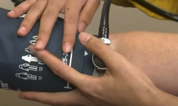
Detection and Diagnosis
The only sure way to detect ocular hypertension is through regular eye exams, which usually include a tonometry test to measure IOP and a visual field test to check for areas of vision loss. Optic nerve imaging may also be done to identify signs of damage.
Conclusion
Ocular hypertension, characterized by elevated intraocular pressure, occurs primarily due to an imbalance in the production and drainage of the eye’s aqueous humor. While it can arise from various causes, such as overproduction of fluid, inadequate drainage, certain medications, or systemic health conditions, its development is strongly influenced by risk factors including age, race, family history, myopia, and diabetes. Although ocular hypertension often exhibits no symptoms, it’s crucial to remember that it can be a significant risk factor for glaucoma. Therefore, regular eye examinations, prompt treatment, and a healthy lifestyle are essential to managing ocular hypertension and maintaining overall eye health.
FAQS
Yes, ocular hypertension is considered a significant risk factor for the development of glaucoma. However, not everyone with ocular hypertension will develop glaucoma.
While there is no cure for ocular hypertension, it can be managed effectively with medication, lifestyle changes, or surgery, if needed. Regular eye exams are crucial to monitor the condition.
Ocular hypertension is often asymptomatic until it progresses to glaucoma. This is why regular eye exams are crucial for early detection and treatment.
Treatment typically involves eye drops to either reduce the production of aqueous humor or improve its drainage. In some cases, laser treatment or surgery may be needed.